Tuesday, 18 August 2020
How labelling brain parts functionally can be overly simplistic: the cerebellum as a case in point

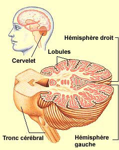
Today I’d like to talk about the cerebellum. To introduce this topic, I’ll remind you that as animals’ bodies evolved and became more complex, they were subjected to greater adaptive pressures to move more and more efficiently, and the cerebellum is a brain structure that was closely involved in this process.
Here’s the most surprising fact about the cerebellum. The human brain as a whole contains about 86 billion neurons. The cerebral cortex accounts contains about 16 billion of these neurons and accounts for about 80% of the brain’s weight. In contrast, the cerebellum accounts for only about 10% of the brain’s weight, but contains nearly 69 billion neurons! Thus more than three-quarters of the neurons in the human brain are located in the cerebellum, even though it is a small structure compared with the brain as a whole.
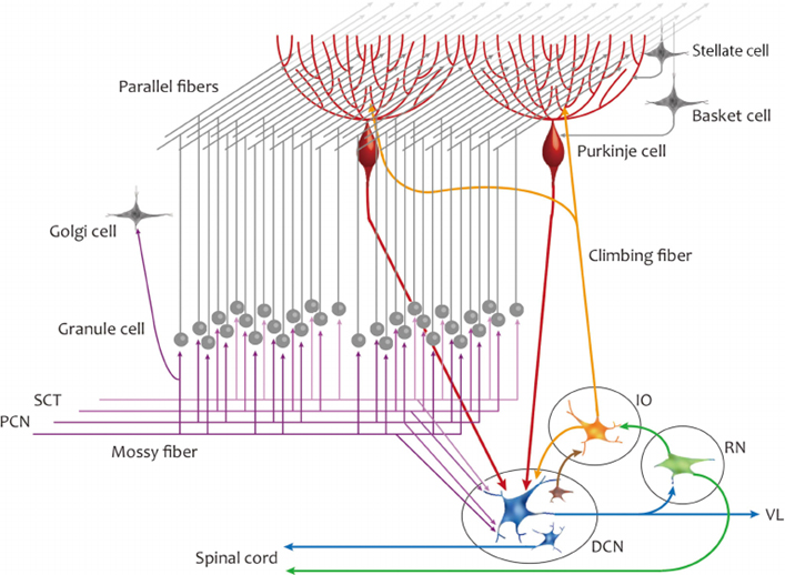
Every signal that enters the cerebellum is processed by some 10,000 of its granule cells, which are among the smallest neurons in the brain (that’s how so many of them can fit in such a small space). But these granule cells are organized into groups of 200,000, within which all of the cells send signals to the same single Purkinje cell (the Purkinje cells are the largest neurons in the cerebellum). This cell in turn relays one signal to the rest of the brain, as shown in the image below (Source: Schematic representation of the neuronal circuit of the cerebellum.).

It has long been known that people with cerebellar lesions have some highly disabling problems with their locomotion or fine motor coordination. The cerebellum was therefore long thought to be involved chiefly in coordinating and synchronizing body movements. But as neuropsychologist Jörn Diedrichsen explains, if you look at the activity of the cerebellum in brain images, you will see that about 70% of its neurons apparently have nothing to do with motor control. Only 30% really activate when the individual is performing body movements. It is now clear that this structure is involved in all of the processes for which we also use the rest of our brain: thoughts, emotions, language and even memory.
Neuroscientists now know that the cerebellum helps to coordinate schemas for numerous types of learning across time (for example, to calculate the right moment to perform a given motor action, in response to a given sensory input). It thus serves as a kind of sub-contractor that can perform this specific kind of calculation on behalf of various mental processes. Consequently, the cerebellum “lights up” in almost all of the tasks observed with brain imaging. Yet its activation often receives less analysis than that of the cortex, which is better known and more highly “valued” in research in general. Diedrichsen revealingly relates that he often receives e-mails from colleagues who ask him why the cerebellum is activating when they are observing a given task, or whether it is not actually really activating and they are instead making errors in their observations.
One example among many others might be this study published in the Journal of Neuroscience in 2017, which showed an activation of certain parts of the cerebellum when the subjects were performing a sentence-completion task. As the authors write, “These results are consistent with a role for the right posterolateral cerebellum beyond motor aspects of language, and suggest that cerebellar internal models of linguistic stimuli support semantic prediction.”
Most of the structures in the brain are in fact composed of numerous sub-regions that maintain specific connections with one another as well as with other brain structures, and the cerebellum is no exception. In its case, it is the most lateral portion, the neo- or ponto-cerebellum which developed the most during hominization, that enables rapid adjustments not only in movements but also in thoughts, and even in emotional reactions (so that they can be appropriate in the eyes of outside observers, which is not always the case following certain injuries to the cerebellum).
Images that arouse emotions activate various areas in the cerebellum, which has direct connections to the amygdala, an important emotional centre in the brain. Moreover, many emotions also have a motor component (for example, fear and its connection with the fight-or-flight reaction). The cerebellum’s tremendous computational capabilities, which are necessary for sensorimotor integration, thus seem to have been used for other neuronal processes in the course of evolution, which represents another example of neuronal recycling.
Body Movement and the Brain | Comments Closed


