


Tuesday, 19 September 2023
Mechanisms underlying anatomical the tracing methods
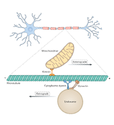 Today I’d like to tell you about the cellular mechanisms underlying the tracing methods used to visualize the paths that axons follow from one structure in the brain to another. The use of these methods in animal models is based on the ability that neurons have to move molecules along the microtubules in their axons. This process is essential to the normal functioning of neurons and is known as axonal transport. It works by means of motor proteins that, when supplied with a bit of energy, constantly change form to drive all kinds of molecules carried in vesicles, a bit like a porter with a load on his back. There are some animations on the Internet that illustrate this process, and it’s pretty impressive!
So when researchers inject certain markers, such as fluorescent proteins, into a part of the brain, they can enter the neurons and be carried along by axonal transport, thus revealing the entire path of the axon, which sometimes runs long distances through various brain structures to its destination. The entire basis of neuroanatomy was established in this way, using anterograde tracing methods, in which the markers are transported from the neuronal cell body to the tip of the axon, and retrograde tracing methods, in which they are carried in the opposite direction.
From the Simple to the Complex | Comments Closed