Thursday, 4 April 2024
The Brain Is Not a Space Shuttle
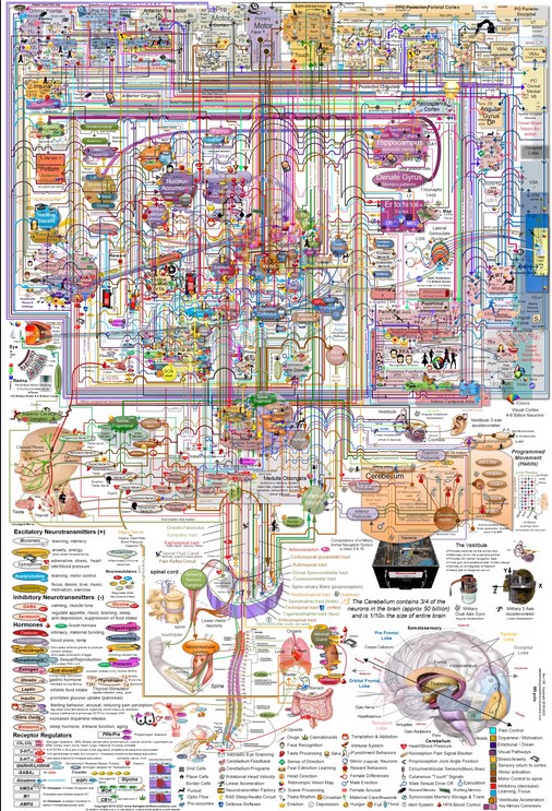
 Recently, someone made me aware of an impressive graphic that attempts to use current neuroanatomical data to show how the brain’s circuits are interconnected, somewhat like the graphics that biochemists use to represent cellular metabolism.
I have never before seen any schematic representation of the brain’s circuits that pulls together so much information, both in its detailed version and in its simplified version, which shows the brain’s main circuits in the sagittal plane. The box in the lower left-hand corner of this graphic states that the research required to develop it was done by an aerospace engineer who had worked on the design of the space shuttle’s guidance system and who spent over four years analyzing over 1000 neuroscientific studies to prep this schematic. (more…)
From the Simple to the Complex | Comments Closed
Friday, 14 July 2017
Metaphors for the Brain’s Anatomy and Functioning

When I’m making presentations about the human brain to live audiences, the quick, easy method I often use to show them a three-dimensional model of a brain synapse is to hold my two fists facing each other, very close together, but not touching. One fist thus represents the axon of the pre-synaptic neuron, while the other represents a dendritic spine on the post-synaptic neuron. This macro model of a synapse is about 20 centimetres long.
In comparison, a real synapse in a mammalian brain is about 1 micron (one thousandth of a millimeter) long. This estimate includes the terminal button (the swelling at the tip of the axon), the dendritic spine (the swelling on a dendrite of the second neuron which receives the connection from the axon of the first), and the synaptic gap (the space between them). Into this gap, the axon of the pre-synaptic neuron releases its neurotransmitters, which immediately bind to the receptors in the membranes of the post-synaptic neuron’s dendritic spine. (more…)
From the Simple to the Complex | No comments


