


Monday, 30 May 2022
Ultrasound localization microscopy provides unprecedented view of blood flow in the brain
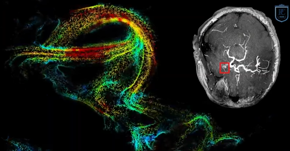 The technologies that can now be used to image various aspects of the anatomy and physiology of the brains of living human beings are triumphs of scientific and technical inventiveness. One of the newest of these techniques is ultrasound localization microscopy, which was recently used to provide the first-ever dynamic images of blood flowing through the capillaries of the brain. This new ability to view blood circulation in the brain so rapidly and precisely opens opportunities for a better understanding of the irrigation of the brain and the problems that can arise with it, such as aneurysms.
In a newspaper article, Chloé Bourquin, a doctoral student in biomedical engineering at Polytechnique Montréal who experiments with using ultrasound localization microscopy to image the human heart, explains this technology as follows:
This method is based on injecting very small air bubbles, as doctors already do to visualize blood flow in echocardiography, another method that uses ultrasound (very high frequency sound waves). Carried along in the blood flow, these bubbles travel throughout the entire body in about 10 minutes, while an echocardiograph records thousands of images of the brain per second, like a camera shooting photographs in high-speed bursts. Powerful calculations are then performed to locate and track each bubble from one image to the next as it flows through the blood vessels. Ultimately, the sum of all these bubbles can be used to produce an extremely detailed map of the blood flow in the brain. This method uses ultrasound to locate the bubbles and thereby visualize the blood vessels on a microscopic scale—hence its name, ultrasound localization microscopy.
Maybe when you read that description, you had the same reaction I did: isn’t it dangerous to inject bubbles into blood vessels? But after a quick search, I found the answer is apparently no, because the total volume of these bubbles is so small: according to one article, less than 200 microlitres in all, making the bubbles about as safe as other contrast agents used in medical imaging. Because that is indeed what these microbubbles do: they serve as targets that the ultrasound waves bounce off of, thereby identifying their locations. In this way, the muddiness of typical echocardiogram images is avoided.
The scientific article describing this advance, entitled Transcranial ultrafast ultrasound localization microscopy of brain vasculature in patients, by Charlie Demené and co-authors, was published in the journal Nature Biomedical Engineering in March 2021. To achieve the resolution of about 25 microns needed to visualize the turbulent blood flow in small aneurysms deep in patients’ brain, the researchers had to find a way of correcting the main possible sources of interference with the ultrasonic signal, such as “micrometric brain-motion artefacts and ultrasonic-wave aberrations induced during transcranial propagation”. As so often happens in getting scientific advances to work properly, the devil was in a detail like this.
So now I’m going to have to add ultrasound localization microscopy to the module on brain-imaging methods on this website. It is a wonderful new tool in the medical imaging arsenal: it is non-invasive, uses no radiation, and costs less than big MRI machines.
Uncategorized | Comments Closed