


Friday, 14 July 2017
Metaphors for the Brain’s Anatomy and Functioning

When I’m making presentations about the human brain to live audiences, the quick, easy method I often use to show them a three-dimensional model of a brain synapse is to hold my two fists facing each other, very close together, but not touching. One fist thus represents the axon of the pre-synaptic neuron, while the other represents a dendritic spine on the post-synaptic neuron. This macro model of a synapse is about 20 centimetres long.
In comparison, a real synapse in a mammalian brain is about 1 micron (one thousandth of a millimeter) long. This estimate includes the terminal button (the swelling at the tip of the axon), the dendritic spine (the swelling on a dendrite of the second neuron which receives the connection from the axon of the first), and the synaptic gap (the space between them). Into this gap, the axon of the pre-synaptic neuron releases its neurotransmitters, which immediately bind to the receptors in the membranes of the post-synaptic neuron’s dendritic spine. (more…)
From the Simple to the Complex | No comments
Monday, 8 August 2016
A Nanometric 3D Representation of a Mouse Cortex Cortex
 The analogy between a real forest and a “forest of neurons” has been drawn many times, but the images produced most recently by the team of Jeff Lichtman and Narayanan Kasthuri (see the first two links below) make it clear yet again that the complexity of the brain’s connections far surpasses that of the densest forest.
As Lichtman has long said, by going down to the scale of the electron microscope and then reconstructing the slightest contacts between axons, dendrites, and neighbouring glial cells slice by slice, we can detect patterns that escape us at the more “macro” scales previously used to model neuronal connectivity. (more…)
From the Simple to the Complex | No comments
Saturday, 18 July 2015
Microscopic Synapses and Giant Microscopes

More and more courses are being offered for free online by prestigious universities. Many of these courses deal with various aspects of the cognitive sciences. One such course is “The Fundamentals of Neuroscience”, from Harvard University (see first link below). This course includes various multimedia features, including an excellent 30-minute documentary video entitled “Connectomics: Big Microscopes & Tiny Synapses” (second link below). This video presents the research being done by Professor Jeff Lichtman and his colleagues in his laboratory, who are using images of real human brains to try to map the connections between their neurons—the synapses. (more…)
From the Simple to the Complex | Comments Closed
Tuesday, 27 May 2014
The Variety and Structural Complexity of Neurons
 The purpose of most of the posts in this blog is to summarize recent studies in the cognitive sciences and attempt to make them more accessible—in particular by providing links to selected pages on this website. But the purpose of some of the other posts is simply to draw attention to existing resources on various aspects of neuroscience. Today’s post falls in the latter category. It deals with the neuron and the work by Kristen Harris and her colleagues to reveal it in all its complexity (see the first two links below). (more…)
From the Simple to the Complex | Comments Closed
Wednesday, 24 July 2013
Oligomers Help Us Keep Our Memories
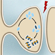 A protein whose molecules have the special ability to stack on top of one another, somewhat like egg cartons, and thus form short, highly stable chains (oligomers), may well play a key role in the mechanisms by which we maintain lasting memories.
Indeed, there is always something magical about remembering something that you haven’t thought about in years. But physically speaking, in what form is the corresponding memory trace maintained in your brain over all that time? Neurobiologists know that the whole process occurs in the synapses (the plastic connections between neurons) and have described molecular mechanisms such as long-term potentiation that partly explain this plasticity. (more…)
Memory and the Brain, Mental Disorders | Comments Closed